Eight bones together comprise our cranial vault. Often it is written in the U.S. (1) that by the age of 25 these bones have fused and that the skull has thus become in essence one large bone. This description is thought based on early (circa 1900) cadaver dissections (2). European anatomy texts and recent Histological examinations of the joints between the cranial bones show that they are dynamic connective tissue structures with a blood supply. Close examination of the joints reveals intentional construction with some joints resembling hinges and other joints resembling sliding guide rails, while others are some combination of the two. It is simple to validate that the cranial bones move for yourself by having someone rest their head in both of your hands while they are lying down. After a few minutes, you should feel a quiet rhythm that is slower than respiration and is the changing pressure of the Cerebral Spinal Fluid pushing against the inside of the skull bones.
How Cranial Bones Jam
Restrictions in the free movement of the cranial bones can originate from many varied sources. Birth trauma, use of suction or forceps, and even C-section deliveries have been linked to cranial lesions. Head injuries often can lead to restrictions. Viral infections have been associated with hardening of the connective tissue that joins the cranial bones. Wearing braces on teeth can have a strong influence on cranial bone function, as well as Dental or cranial surgery. An interesting source is Caesarian section delivery. The thinking here is that the abrupt pressure change from the womb to the outside world is too quick for the infant’s cranium to adapt to and the cranial bones jam(3). Also, traveling through the birth canal initiates the proper function of the cranium. Whatever the source, the consequence is felt as inhibition of movement in the bones. If there is a lot of restriction, these skulls have a harder, more solid, less alive feel than a properly functioning cranium and it becomes quite easy to appreciate how this would influence health and vitality. Often areas that are inhibited are easy to identify, and when those bones start working together, that change is also easy to feel.
Why the Cranium is Assembled Using Pliable Joints
The cranium” ‘s ability to manage trauma is greatly enhanced by this flexibility. Shock loads that would crack a fused skull like an egg are distributed around the cranium by this flexion, as in a Tensegrity structure, or a Turtle shell. It is also pleasing to note the similarity in design between the cranium and the Tectonic Plates of our planet. Another of the primary functions of cranial motion is to facilitate the flow of Cerebral Spinal Fluid (CSF) around the brain, flushing toxins and metabolic wastes out of the central nervous system. The cranium supports this flow by altering its shape as the internal pressure of the CSF changes. Inside the cranium the pressure of the CSF increases until valves open, releasing the CSF back into the bloodstream and lowering the pressure. It is thought that these pressure changes assist the CSF in penetrating the nooks and crannies of the brain, working as a form of pump for the movement of the fluid. If the bones of the cranium flex properly the skull adapts to the changing pressure of the CSF, slowing the rate of change, like a river broaching its banks. This increases the length of time before the valves controlling the release of CSF reach the trip point pressure and open, generating a longer cycle and preventing sharp pressure changes and the resulting stress on the tissues of the brain.
How Restrictions Influence the Flow of Cerebral Spinal Fluid
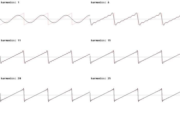
The more restricted the movement of the cranial bones the more the flow of CSF will be restricted(2). An interesting analogy to demonstrate this phenomenon is that of a river basin. In its natural configuration as the height of a river increases, there is a corresponding increase in width as the river breaches its banks and floods the surrounding land(expansion of the cranium). Because its flow is not restricted the water velocity and pressure change gradually and it is less likely that there will be hard spikes in the rate of flow. If the surrounding land is isolated from the river by dikes or levies, pressure swings are rapid, and velocity changes quickly, and consequently much more destructive. In engineering, we would describe the difference using waveforms. The normal rhythm would look like a sine wave, and the restricted cranium waveform would be a sawtooth.
Sine Wave Transitioning to a Saw Tooth Waveform (From Wikipedia)
The Harmonics are additional energy contained in the wave. In the cranium, the wavelength also shortens, again increasing the amount of energy the cranial system has to dissipate. You can imagine the effect of CranioSacral Therapy as this illustration runs backward, from sawtooth to sine.
Consequences of Restrictions in the Flow of Cerebral Spinal Fluid
Fast changes in CSF pressure can result in many different physical, cognitive, and emotional challenges. It is as if the fast changes put the brain into a defensive mode where it will only minimally function as if to protect its core from harsh changes. Hypervigilant protection is a hallmark of a hard head. As the CSF pressure change rate slows down, the brain appears to be willing to begin to explore more of its potential, moving from a defensive into a learning mode. Behaviors as fundamental as communication or holding a spoon may improve. Genetic and congenital disorders as difficult as Autism and CP, which also include hardheadedness usually will improve by eliminating restrictions in cranial function. It is difficult to relax and take in the world when your head is pounding.
Brain Stem and Cranial Joint Alignment
The bone at the base of the skull, the Occiput, is often thought of as the first vertebrae of the cervical spine. As discussed above, occiput alignment is influenced by the relationship of the bones of the cranial cavity and other bones of the skull. In turn, the occiput influences the orientation of the atlas, the first anatomical cervical vertebrae. This joint is considered by many to be of prime importance in postural alignment and also in its influence on brain stem function. The brain stem begins at this joint. Consequently, any misalignment or tension here may compromise neuron function. Atlas adjustments can immediately be experienced as a greater sense of calm, diffuse anxiety, and greater clarity of thought. The occiput/atlas joint consists of two saddle joints to make a stable platform for anterior/ posterior skull motion, as in nodding yes. There is some trade-off with flexibility and the soft tissue can become quite dense around the joint, restricting movement. Corrections usually involve setting the alignment and allowing the body to relearn the proper orientation to this alignment. This may take many repetitions over a long period before it “sticks”. Improving the efficacy and efficiency of atlas adjustments is one of the primary developmental goals of Manual Therapy research.
How to Correct Cranial Restrictions
Corrections to cranial bone function can be done in a variety of ways. It is often faster to work below the amount of force that causes the bones to lock up, which can be quite minimal. Corrections can take only a few minutes, to focused work over several sessions, to many years if at all. The results are experienced as greater freedom in the physical, emotional, and cognitive domains.

